
When you’re dealing with back pain, getting an accurate diagnosis is the first step toward finding relief. Doctors frequently use two imaging tools to look inside the spine: X-rays and MRIs.
While both provide a look inside the body, they capture entirely different things. Knowing when each is necessary can help you understand your diagnostic process.
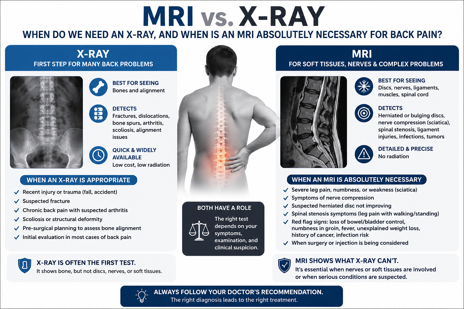
The Core Difference: Bones vs. Soft Tissue
The main distinction between these two imaging technologies comes down to what they can actually see:
X-Ray (Radiography): Uses a small amount of radiation to capture high-contrast images of dense materials. It is excellent for viewing bone structure but largely misses soft tissues.
MRI(Magnetic Resonance Imaging): Uses powerful magnets and radio waves (no radiation) to create highly detailed, cross-sectional images of soft tissues like muscles, nerves, ligaments, and spinal discs.
When Do You Need an X-Ray?
An X-ray is almost always the first line of defense when investigating back pain. Because it is quick, widely available, and cost-effective, doctors use it to rule out structural or skeletal issues.
Common Reasons for a Spinal X-Ray:
Recent Trauma: Checking for fractures or dislocations after a fall, car accident, or sports injury.
Postural or Structural Alignment: Identifying conditions like scoliosis (abnormal curvature of the spine) or kyphosis.
Spinal Instability: Spotting conditions like spondylolisthesis, where one vertebra slips forward over another.
Age-Related Bone Wear: Evaluating the spine for osteoarthritis or bone spurs (osteophytes).
If your back pain is relatively new and isn’t accompanied by severe neurological symptoms, a clear X-ray combined with a physical exam is often enough to start a conservative treatment plan, such as physical therapy.
When is an MRI Absolutely Necessary?
An X-ray only tells half the story. If your bones look perfectly healthy but you are still in severe pain, the culprit is likely a soft tissue issue. This is where an MRI becomes essential.
Common Reasons for a Spinal MRI:
Suspected Disc Issues: Identifying herniated, bulging, or degenerative discs that don’t show up on a standard X-ray.
Nerve Compression or Sciatica: If you experience shooting pain, numbness, tingling, or weakness traveling down your leg, an MRI can pinpoint exactly which nerve root is being pinched.
Spinal Stenosis: Visualizing the narrowing of the spinal canal, which puts pressure on the spinal cord.
Infections or Tumors: Detecting rare but serious underlying causes of back pain, such as spinal tumors, cysts, or deep tissue infections.
Comparing the Diagnostic Tools
Feature X-Ray MRI Primary Target Bones and skeletal alignment Soft tissues, discs, and nerves Imaging Method Low-dose ionizing radiation Magnetic fields and radio waves Scan Duration 5 to 10 minutes 30 to 60 minutes Main Advantage Fast, inexpensive, excellent for bone fractures Incredibly detailed view of nerves and discs Main Limitation Cannot see discs, muscles, or nerve pinching Higher cost, requires staying perfectly still in a closed space