
Revolutionizing Spine Care: A Comprehensive Guide to Unilateral Biportal Endoscopic (UBE) Surgery
Back pain and degenerative spine conditions are among the leading causes of disability worldwide. For decades, traditional open spine surgeries—while effective—came with significant drawbacks: large incisions, extensive muscle stripping, prolonged hospital stays, and lengthy recovery periods.
In recent years, minimally invasive spine surgery (MISS) has transformed the orthopedic and neurosurgical landscape. Among the most cutting-edge and rapidly evolving techniques is Unilateral Biportal Endoscopic (UBE) Surgery. Offering unprecedented visualization, precision, and faster recovery times, UBE is quickly becoming a gold standard for treating various spinal pathologies
Understanding UBE Surgery: What Is It?
Unilateral Biportal Endoscopic spine surgery is an advanced form of minimally invasive spine surgery. The term itself breaks down the core mechanics of the procedure:
Unilateral: The entire surgery is performed through one side of the back, even if the condition affects both sides (using specialized “over-the-top” decompression techniques).
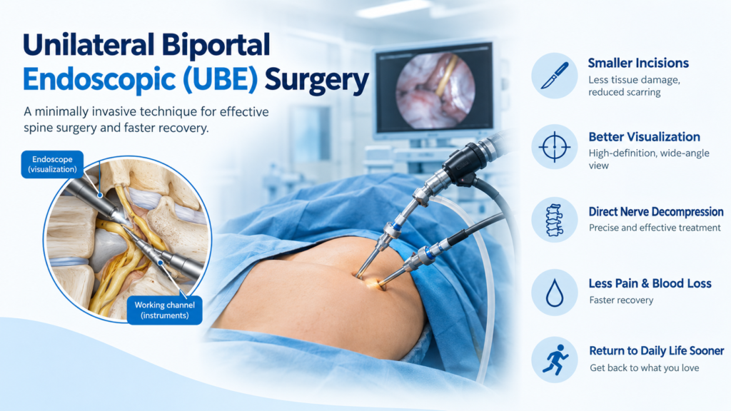
Biportal: Unlike traditional single-port endoscopes where the camera and instruments share the same channel, UBE utilizes two separate portals (small incisions, usually around 5 mm each).
Portal 1 (Viewing Portal): Dedicated solely to the high-definition endoscope, providing a crystal-clear, magnified view of the surgical field.
Portal 2 (Working Portal): Dedicated to standard arthroscopic and specialized spinal instruments, allowing the surgeon to cut, grab, shave, and decompress with high dexterity.
How UBE Differs from Traditional and Other Minimally Invasive Methods
| Feature | Open Spine Surgery | Microendoscopic Surgery (tubular/single-port) | UBE Surgery |
| Incision Size | Large (5 to 10+ cm) | Medium (1.5 to 2.5 cm tubular retractor) | Two tiny punctures (approx. 0.5 cm each) |
| Muscle Damage | Extensive detachment and retraction | Moderate (tube dilation pushes muscle aside) | Minimal (utilizes continuous saline fluid flow and independent tool movement) |
| Visualization | Direct naked-eye view | Limited by narrow tubular diameter | Wide panoramic view with high-definition magnification |
| Instrument Maneuverability | High | Restricted by the confines of a single tube | High (instruments move independently of the camera) |
| Hospital Stay | 3 to 7 days | 1 to 2 days | Same-day discharge or 1-day stay |
Conditions Treated with UBE Surgery
UBE is remarkably versatile. Spine surgeons utilize this technique to treat a wide array of degenerative and compressive spine disorders, primarily in the lumbar (lower) spine, though cervical (neck) and thoracic applications are growing. Common indications include:
Lumbar Disc Herniation (Slip Disc): Removal of the herniated portion of the disc that is pressing on the nerve root, relieving sciatica and radiating leg pain.
Lumbar Spinal Stenosis: Decompression of narrowed spinal canals by removing thickened ligamentum flavum and overgrown bone spurs (osteophytes).
Foraminal Stenosis: Widening the neural foramina (the bony openings where nerves exit the spine) to alleviate chronic nerve root compression.
Synovial Cysts: Removal of fluid-filled cysts that press against the spinal nerves.
1. Preparation and Anesthesia
The patient is positioned prone (face down) on a specialized surgical table to minimize abdominal pressure and reduce bleeding. The procedure is typically performed under general anesthesia, though regional or epidural anesthesia may be used in select cases.
2. Portal Creation
The surgeon identifies the target spinal level using fluoroscopy (real-time X-ray). Two tiny incisions (about 5 mm each) are made on one side of the spine.
The upper incision accommodates the endoscope.
The lower incision serves as the working channel.
3. Continuous Irrigation
Unlike traditional surgery where visibility relies on air and electrocautery smoke clearance, UBE is performed underwater. A constant stream of sterile saline solution flows through the working area. This serves three vital purposes:
Keeps the surgical field impeccably clean by washing away blood.
Provides natural hydrostatic pressure to control bleeding from tiny blood vessels.
Magnifies and clarifies the anatomical structures under the endoscope.
4. Decompression
Using specialized miniature high-speed drills, punches, and micro-curettes inserted through the working portal, the surgeon carefully removes the offending bone spurs, thick ligaments, or herniated disc material. The endoscope provides a 4K-magnified view, allowing the surgeon to distinguish nerve tissues from surrounding structures with extreme precision.
5. Closure
Once adequate decompression is verified and hemostasis is achieved, the saline is drained, the instruments are removed, and the tiny skin incisions are closed with skin glue or a single stitch. No bulky drains or deep muscle sutures are typically required.
Advantages of UBE Surgery
The clinical benefits of UBE over older surgical methodologies are profound:
Preservation of Spinal Stability: Because muscles are not stripped off the bone and the posterior tension band (spinous processes and ligaments) remains largely intact, the natural biomechanical stability of the spine is preserved.
Superior Visualization: The independent movement of the camera and instruments, combined with water-medium magnification, allows surgeons to see corners and recesses of the spinal canal that are hidden in tubular or open surgeries.
Reduced Postoperative Pain: Minimized soft-tissue trauma translates directly to significantly less post-surgical pain and a dramatic drop in the need for narcotic painkillers.
Faster Functional Recovery: Patients are frequently encouraged to walk within hours of the procedure. Return to light daily activities and work happens weeks sooner than with open procedures.
Cosmetic Appeal: The two small puncture marks heal into barely noticeable scars.
Recovery and Rehabilitation
Recovery following UBE surgery is remarkably swift compared to traditional open back surgeries:
Day of Surgery: Most patients stand and walk a few hours post-operation under the guidance of a physical therapist. Many are discharged home the same day or the following morning.
Weeks 1 to 3: Light indoor activities are encouraged. Patients should avoid heavy lifting (anything over 5 kg), prolonged sitting, and excessive twisting or bending.
Weeks 4 to 6: Supervised physical therapy focusing on core strengthening, posture correction, and gentle stretching begins.
Month 3 and Beyond: Most patients can resume normal physical activities, recreational sports, and strenuous work duties, provided they maintain proper ergonomics.
Risks and Considerations
While UBE is exceptionally safe and effective, it is still a surgical procedure on the spine. Potential risks—though rare in the hands of an experienced, fellowship-trained endoscopic spine surgeon—include:
Dural tear (leakage of spinal fluid)
Transient nerve irritation or numbness
Superficial wound infection
Incomplete decompression or recurrence of disc herniation
Choosing a surgeon with specialized training in endoscopic spine techniques is the single most critical factor in minimizing these risks.
Conclusion
Unilateral Biportal Endoscopic (UBE) surgery represents a monumental leap forward in spinal care. By merging the precision of endoscopy with the dexterity of independent biportal instrumentation, it grants surgeons the ability to treat complex spinal pathologies with minimal collateral damage to healthy tissues. For patients suffering from debilitating back and leg pain, UBE offers a reliable path toward rapid healing, less pain, and a swift return to a vibrant, active lifestyle.