
Revolutionizing the Operating Room: The Power of O-Arm and Intraoperative 3D Imaging Systems
For decades, modern surgery has relied heavily on the surgeon’s skill, tactile feedback, and conventional 2D imaging tools like standard fluoroscopy. While helpful, 2D images often flatly represent complex three-dimensional anatomies, sometimes leaving critical details to guesswork or requiring postoperative scans that can lead to unexpected revision surgeries.
Enter the O-Arm and intraoperative 3D imaging systems—a groundbreaking technological leap that is transforming complex spine, orthopedic, and cranial surgeries.
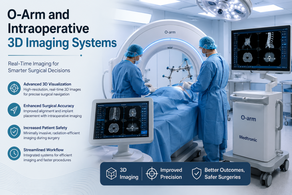
What is an O-Arm System?
The O-Arm is a mobile, multidimensional surgical imaging device—most notably developed by Medtronic—designed specifically for use directly inside the operating theater.
True to its name, the device features a mobile gantry shaped like an “O”. During surgery, the open gantry can be wheeled into position and closed electronically to form a complete 360-degree ring around the patient. It provides:
Real-time 2D fluoroscopy
High-definition 3D volumetric multi-planar imaging (axial, sagittal, and coronal planes) akin to a diagnostic CT scan, but right on the operating table
How Intraoperative 3D Imaging Works
Unlike traditional workflows that require pre-operative scans and separate post-operative evaluations, the O-Arm bridges the gap entirely.
Intraoperative Scanning: When a surgeon reaches a critical phase of an operation—such as placing pedicle screws in the spine—the O-Arm is positioned over the surgical site. It rotates to capture a 3D scan in mere seconds.
Navigation Integration: The system seamlessly integrates with surgical navigation platforms (like StealthStation™). It acts like an advanced GPS for the human body, automatically registering patient data so the surgeon can track instruments in real-time.
Immediate Verification: The surgeon can instantly verify if an implant or screw is in its absolute optimal position. If an adjustment is needed, it is corrected before the patient leaves the operating room, drastically cutting down the necessity of secondary revision
Key Benefits of O-Arm Technology
Unmatched Precision and Safety: In delicate procedures—such as spinal fusion, tumor removals, or instrumentation near the spinal cord and major blood vessels—accuracy is paramount. O-Arm imaging elevates placement accuracy significantly, minimizing the risk of nerve or vascular injury.
Elimination of Secondary Surgeries: Studies show that traditional 2D methods can occasionally result in misplaced hardware only discovered on post-op scans. Intraoperative 3D imaging allows teams to catch and fix these issues immediately, sparing the patient a second trip to the operating room.
Optimized Workflow and Efficiency: By eliminating the need to transfer patients to radiology mid-procedure or schedule follow-up diagnostic CTs, the O-Arm streamlines the entire surgical timeline.
Minimally Invasive Surgery (MIS) Support: Enhanced visualization allows surgeons to perform complex procedures through smaller incisions, leading to less blood loss, reduced risk of infection, and faster patient recovery.
Adjustable Radiation Control: Modern systems are built with patient and staff safety in mind, featuring low-dose imaging protocols that align with the ALARA (As Low As Reasonably Achievable) principle.
The Future of the Operating Room
The integration of O-Arm technology alongside surgical robotics and advanced navigation represents a golden standard in modern medicine. By replacing uncertainty with crystal-clear, real-time 3D visualization, these systems give medical teams supreme confidence and offer patients safer procedures with better long-term outcomes.
As hospitals continue to adopt these smart operating suites, the boundary between diagnostic imaging and surgical execution continues to blur—paving the way for a safer, more precise surgical future.