
Chronic lower back pain remains one of the leading causes of disability worldwide. For decades, the medical consensus surrounding Intervertebral Disc Degeneration (IVDD) accepted a frustrating reality: once a spinal disc begins to break down, the damage is irreversible. Traditional management strategies—ranging from physical therapy and epidural steroid injections to radical surgical interventions like spinal fusion—focus almost entirely on masking symptoms or stabilizing a structurally compromised spine.
However, a fundamental paradigm shift is underway in orthopedics and regenerative medicine. Rather than managing end-stage structural decay, researchers and clinicians are deploying biologics and stem cell therapies aimed at halting—and potentially reversing—the cellular cascade of disc degeneration.
Understanding the Enemy: The Pathophysiology of Disc Degeneration
The intervertebral disc consists of three integrated structures:
Nucleus Pulposus (NP): The gelatinous core rich in proteoglycans and type II collagen, responsible for absorbing shock and maintaining fluid retention.
Annulus Fibrosus (AF): The tough, fibrous outer ring composed of concentric collagen bands that contain the inner nucleus.
Cartilaginous Endplates (CEPs): Thin layers of hyaline cartilage separating the disc from adjacent vertebral bodies.
The health of this complex system hinges on extracellular matrix (ECM) homeostasis—a delicate balance between matrix synthesis and breakdown. The adult intervertebral disc is the largest avascular tissue in the human body. It relies entirely on passive diffusion from microvessels in the vertebral endplates for nutrient supply and waste removal.
When degeneration begins, this transport route degrades. The disc environment shifts into an avascular, hypoxic, hyperosmotic, acidic, and low-nutrient microenvironment. Native NP cells undergo premature senescence and apoptosis, triggering a shift toward catabolism. Pro-inflammatory cytokines (such as TNF-$\alpha$, IL-1$\beta$, and IL-6) upregulate matrix metalloproteinases (MMPs) and ADAMTS enzymes, breaking down collagen type II and aggrecan. As proteoglycan concentration drops, the disc loses osmotic pressure, dehydrates, and collapses, leading to mechanical instability and discogenic pain.
Biologics: Re-Balancing the Anabolic-Catabolic Axis
Biological therapies introduce bioactive substances directly into the degenerated disc to quell inflammation and reactivate cellular matrix production
1. Growth Factor Injections
Growth factor therapy aims to tip the local microenvironment back toward matrix synthesis. Promising agents include:
Bone Morphogenetic Proteins (BMP-2, BMP-7 / OP-1): Potent stimulators of extracellular matrix synthesis that increase proteoglycan and collagen production in nucleus pulposus cells.
Growth and Differentiation Factor-5 (GDF-5 / BMP-14): Crucial for disc development, GDF-5 promotes NP cell proliferation and ECM regeneration.
Transforming Growth Factor-Beta (TGF-$\beta$): Synergizes with other molecules to drive chondrogenic differentiation and matrix deposition.
While direct growth factor injections yield immediate metabolic shifts, their clinical utility as monotherapies is limited by short biological half-lives, often requiring repeated high-dose administrations that carry a risk of unwanted tissue calcification
2. Autologous Biologics: PRP and Bone Marrow Aspirate Concentrate (BMAC) Platelet-Rich Plasma (PRP) and BMAC leverage autologous, multi-factorial cocktails:
PRP delivers concentrated platelets that release growth factors (PDGF, TGF-$\beta$, IGF-1, VEGF) upon degranulation. Intradiscal PRP injections have shown significant utility in early-stage disc disease by reducing local inflammation and promoting cell survival.
BMAC offers a dual mechanism: a native population of nucleated stem cells combined with an anti-inflammatory protein profile (such as IL-1 receptor antagonist, IL-1RA).
Stem Cell Therapy: Replenishing cellular machinery
When disc degeneration progresses to moderate stages, the resident population of viable NP cells drops dramatically, rendering growth factor therapy alone insufficient. Stem cell transplantation introduces fresh, responsive cellular machinery to rebuild damaged matrix architecture.
Primary Cell Sources for Disc Regeneration
| Cell Type | Advantages | Challenges |
| Bone Marrow Mesenchymal Stem Cells (BMSCs) | • Extensively studied • Robust chondrogenic potential • Proven paracrine anti-inflammatory effects | • Requires invasive aspiration • Yield declines with patient age • Survival under extreme hypoxia is challenging |
| Adipose-Derived Stem Cells (ADSCs) | • High cell yield via lipoaspirate • Easy harvesting profile • Strong immunomodulatory capability | • Variable differentiation capacity • Sensitive to acidic microenvironments |
| Umbilical Cord MSCs (UC-MSCs) | • Allogeneic, off-the-shelf potential • Low immunogenicity • High proliferation rate | • Regulatory constraints • Donor-to-donor batch variability |
| Endogenous NP Progenitors (ProNPs) | • Pre-adapted to the harsh disc environment • Directly committed to NP phenotype | • Extremely limited harvest quantities • Difficult to expand in vitro |
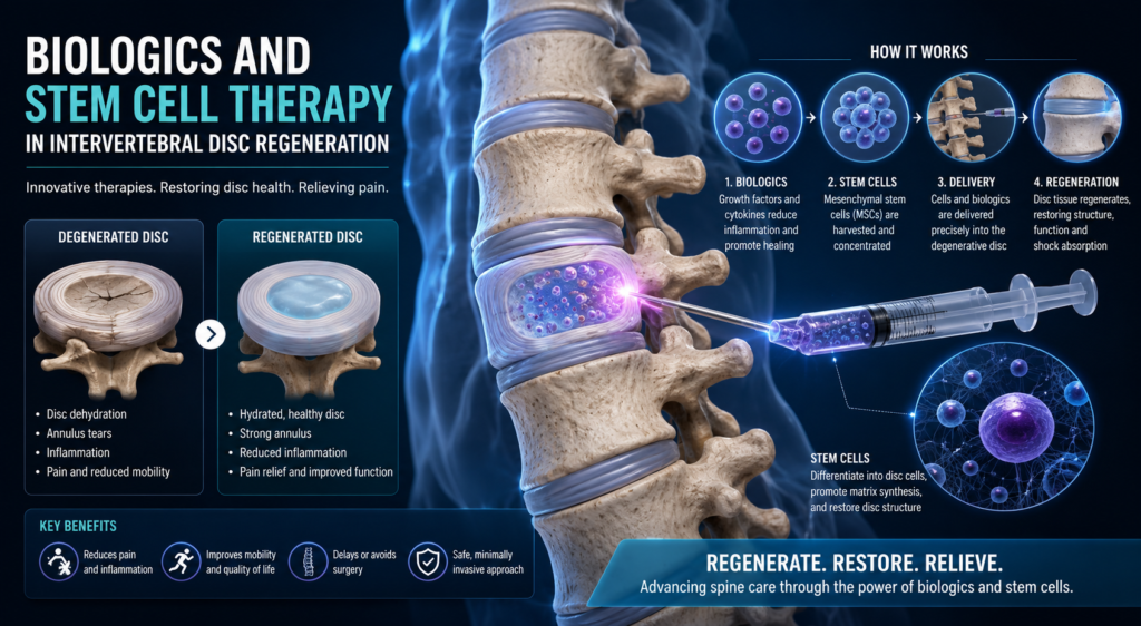
Mechanisms of Stem Cell Action: Direct vs. Paracrine
Initial stem cell strategies assumed transplanted cells would directly engraft and transdifferentiate into functional nucleus pulposus-like cells. While direct differentiation does occur, research shows the dominant therapeutic driver is paracrine signaling:
Paracrine Factor Secretion: MSCs act as local cellular bio-factories, releasing secretomes filled with IGF-1, TGF-$\beta$, anti-inflammatory cytokines (IL-10, IL-1RA), and tissue inhibitors of metalloproteinases (TIMPs).
Matrix Rescue: These signals wake up surrounding native NP cells, stimulating them to resume proteoglycan synthesis while blocking inflammatory cascades.
Exosome Signaling: MSCs release extracellular vesicles (exosomes) loaded with microRNAs and proteins that suppress local cell apoptosis and mitigate oxidative stress.
Navigating the Microenvironment: Biomaterials and Scaffolds
Injecting bare stem cells or growth factors directly into a degenerated disc presents major hurdles: cells leak through the needle track, and those that remain often die due to the acidic, hypoxic, and mechanical stresses inside the disc.
Hydrogels (Hyaluronic Acid, Alginate, Chitosan, Collagen): Hydrogels mimic the native nucleus pulposus matrix, providing structural support, retaining moisture, and protecting transplanted cells from mechanical shear forces during spinal load-bearing.
Controlled-Release Microspheres: Polymer carriers (like PLGA) release growth factors slowly over weeks or months, avoiding early bursts and ensuring sustained biological stimulation.
Translation to the Clinic: Progress and Challenges
Clinical trials evaluating MSC and biologic injections for discogenic pain consistently demonstrate statistically significant improvements in Visual Analog Scale (VAS) pain scores and Oswestry Disability Index (ODI) outcomes. MRI follow-ups frequently show stabilized or improved T2-signal intensity, reflecting preserved fluid content within treated discs.
Despite these encouraging results, several major hurdles must be cleared before these therapies become standard clinical practice:
Hostile Environment Adaptability: Degenerated discs lack vascularization, making oxygen and nutrient availability extremely sparse. Pre-conditioning MSCs under hypoxic conditions or genetically modifying cells to overexpress survival factors are key strategies currently being explored.
Standardization and Dosing: Clinical trials vary widely in cell dosing (ranging from 1 million to over 20 million MSCs per disc), delivery vehicles, and patient selection criteria.
Patient Stratification: Biologics and stem cell therapies require a viable cellular population to work with. They are most effective in Pfirrmann Grade II to IV degeneration (mild-to-moderate loss of disc height with intact annulus fibrosus). Patients with complete disc collapse (Pfirrmann Grade V) or massive structural herniation remain candidates for surgical reconstruction rather than biologics.
The Path Ahead
The future of spine care is shifting from mechanical management toward biological preservation. Advances in CRISPR gene editing, stem-cell-derived exosome isolations, and smart biomimetic hydrogels are addressing the physical and biological challenges of the intervertebral disc.
By treating the underlying biological degradation rather than simply managing chronic pain, biologics and stem cell therapies are establishing a new frontier in orthopedics—one where structural restoration and true biological tissue regeneration are becoming achievable clinical realities.