
Severe spinal deformities
such as high-degree scoliosis, severe kyphosis, or revision cases with distorted anatomy—present some of the highest-stakes challenges in orthopedic and neurological surgery. Correcting a spine that rotates, twists, or buckles requires millimetric precision. One minor deviation during pedicle screw insertion or alignment correction can lead to nerve damage, vascular injury, or structural failure.
Historically, surgeons relied on open exposure, tactile feel, freehand techniques, and repeated 2D X-ray fluoroscopy. Later came external 3D navigation monitors and robotic assistance. While these tools improved accuracy, they introduced a common surgical challenge: attention shift. Surgeons had to look away from the patient’s actual spine to reference a monitor across the room, mentally bridging the gap between digital imaging and physical anatomy.
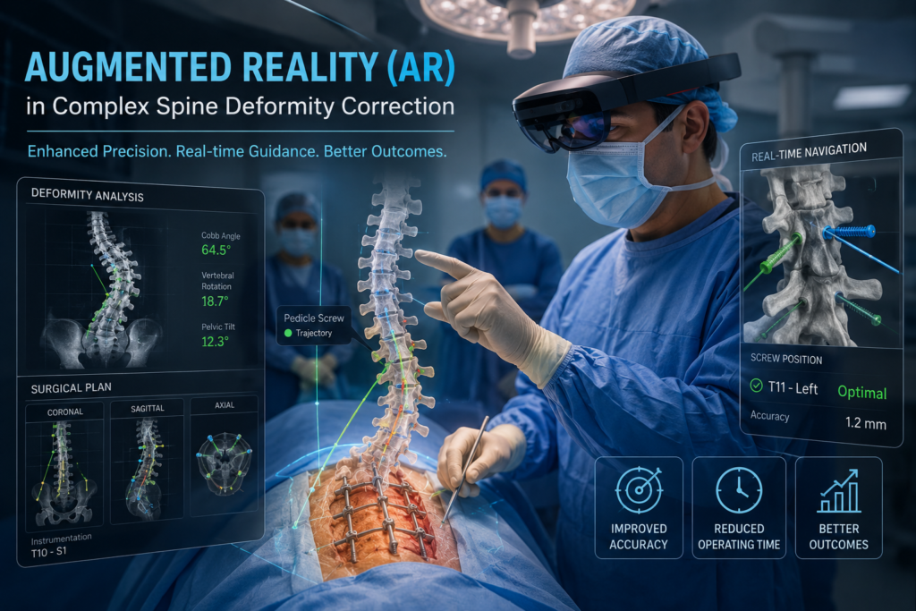
Augmented Reality (AR) eliminates this disconnect by projecting 3D anatomical data directly onto the surgical field through head-mounted displays (HMDs) or surgical visors. The result is equivalent to “X-ray vision,” allowing surgeons to see hidden sub-surface bone structures, trajectory paths, and vital organs without taking their eyes off the patient
The Core Challenge: Why Complex Deformities Need High Precision
Spine deformity correction is not simply about fixing a straight column; it is a dynamic, 3D structural reconstruction. In complex scoliosis or revision cases:
Distorted Anatomy: Normal bone landmarks are often missing, fused from prior operations, or severely rotated.
Tight Safety Margins: Pedicle corridors—the bony bridges where anchoring screws are inserted—can be as narrow as 2 to 4 millimeters. The spinal cord and major blood vessels sit directly alongside these corridors.
Rigid vs. Mobile Segments: Deformities require multiple anchors across dozens of spinal segments to safely distribute corrective forces.
Traditional freehand techniques carry malposition rates estimated between 10% and 15% in complex cases. AR navigation directly targets this margin of error.
How AR Works in the Operating Room
| Step | Process | Clinical Value |
| 1. 3D Mapping | High-resolution preoperative CT or intraoperative 3D scans construct a digital patient spine model. | Establishes patient-specific anatomy, hardware trajectories, and screw dimensions. |
| 2. Registration | Optical trackers align the digital 3D model with the physical spine on the operating table. | Ensures sub-millimeter correlation between projected holograms and real tissue. |
| 3. Real-Time Overlay | Through an AR visor (e.g., FDA-cleared systems like Xvision), 3D anatomical overlays project onto the surgeon’s direct line of sight. | Preserves direct line-of-sight visual feedback, eliminating ergonomic strain and distraction. |
Key Clinical Advantages in Deformity Correction
1. Superior Pedicle Screw Accuracy
Clinical studies on AR-assisted spine surgery consistently demonstrate screw placement accuracy rates ranging between 94% and 99%. In complex scoliosis, where dysplastic or rotated pedicles increase breach risks, AR provides high-precision visual confirmation before bone drilling begins.
2. Elimination of Attention Shift & Improved Ergonomics
Conventional navigation forces a surgeon to look up at external screens to verify position while operating lower down. AR preserves direct line of sight. Maintaining sight on the physical surgical site reduces cognitive load, minimizes micro-movements, and speeds up complex multi-level instrumentation.
3. Reduced Radiation Exposure
Standard multi-level deformity operations traditionally require frequent intraoperative fluoroscopy (X-rays) to confirm screw trajectory and curve alignment. By providing real-time 3D optical tracking, AR significantly reduces the reliance on repeated live X-rays, protecting both the patient and operating room staff from cumulative radiation.
4. Supporting Minimally Invasive Surgery (MIS)
In selected adult spinal deformities, less invasive approaches cut down on blood loss and recovery times. Because MIS limits direct anatomical exposure, AR fills the vision gap by rendering sub-surface structures transparently through the skin and soft tissue.
Current Limitations & Roadblocks to Widespread Adoption
Despite its benefits, AR adoption in routine spine surgery faces several hurdles:
Cost and Infrastructure: High capital expenditure for AR headsets, optical tracking camera systems, and surgical integration remains a barrier for community hospitals.
Soft Tissue Shift & Deformity Reduction: During complex deformity correction, once a surgeon applies force to reduce a spinal curve or performs an osteotomy, the relative positioning of individual vertebrae changes. AR systems must continuously re-register or track individual dynamic segments to maintain sub-millimeter precision.
Headset Ergonomics: While headset design is improving, current hardware adds weight and can cause surgeon fatigue during long 6 to 10 hour deformity cases.
Learning Curve: Transitioning from traditional tactile navigation or external monitor workflows to head-mounted holographic interfaces requires dedicated surgical training.
The Horizon: AI, Robotics, and Beyond
The future of complex deformity surgery lies in converging AR, Artificial Intelligence (AI), and Surgical Robotics.
Upcoming AR platforms aim to incorporate predictive AI models that display real-time mechanical stress overlays. Surgeons will be able to project pre-bent correction rods virtually onto the curved spine, simulating biomechanical forces, junctional breakdown risks, and alignment goals before making physical cuts or placing permanent implants. When paired with robotic arms for drilling execution, AR will serve as the primary visual control center for the surgical team.
Conclusion
Augmented Reality is moving spine deformity correction away from surgical estimation toward digitally verified precision. By projecting dynamic 3D imaging directly into the surgeon’s line of vision, AR improves accuracy, protects critical neurovascular structures, reduces radiation, and streamlines complex reconstruction. As technology lowers hardware costs and refines multi-segment tracking, AR is set to become a core standard of care in complex spinal reconstruction.