
Core Concepts & Technology Integration
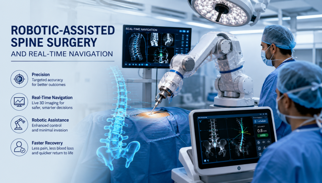
Definition & Purpose: Robotic-assisted spine surgery combines rigid robotic arms with 3D computer navigation to assist surgeons in planning trajectories and accurately placing hardware.
Real-Time Navigation Mechanics: Infrared optical cameras or electromagnetic trackers map surgical instruments in real time against preoperative or intraoperative 3D imaging.
Rigid Frame Alignment: Dynamic reference frames attached to the patient’s spine ensure the navigation system updates instantly if the patient moves.
Preoperative Planning Software: Surgeons create customized 3D trajectory plans for each screw, optimizing size, length, and angle based on high-resolution CT scans.
Clinical & Patient Benefits
5. High Pedicle Screw Accuracy: Precision rates for pedicle screw placement frequently exceed 95–98%, significantly reducing the risk of spinal cord or blood vessel breach. 6. Lower Radiation Exposure: By shifting reliance away from continuous 2D fluoroscopy, both operating room staff and patients experience substantially reduced intraoperative radiation. 7. Minimally Invasive Surgery (MIS): Enables smaller incisions, preserving surrounding paraspinal muscle tissue and soft structures. 8. Faster Recovery Times: Reduced soft-tissue disruption translates to decreased intraoperative blood loss, less pain, and shorter hospital stays. 9. Reduced Revision Surgery Rates: Superior initial placement accuracy reduces the need for secondary surgeries due to misplaced hardware
Surgical Applications & Workflow
10. Complex Deformity Correction: Crucial for challenging cases like severe scoliosis, kyphosis, and revision surgeries where anatomical landmarks are altered. 11. Intraoperative 3D Imaging: Systems integrate with devices like the O-arm or intraoperative CT (iCT) to confirm implant placement before closing the incision. 12. Shared Control (Robotic Arm Guidance): The robot positions a rigid arm guide along the planned trajectory, while the surgeon maintains manual control to drill and place the screw. 13. Tactile Feedback Limitations: Most current spine robots lack haptic feedback, requiring surgeons to rely heavily on visual cues and real-time navigation displays.
Challenges & Limitations
14. High Capital Acquisition Costs: Initial procurement of robotic units and navigation towers can range from $800,000 to over $1.5 million, plus ongoing maintenance fees. 15. Steep Learning Curve: Surgical teams require dedicated training to adapt to registration protocols, platform software, and workflow changes. 16. Risk of Registration Drift: Skydiver or patient movements, or bumping the reference array, can cause small misalignment errors between real-time positioning and visual maps. 17. Skiving Risks: When drilling into steep anatomical angles, soft tissue pressure or drill bits sliding off bone (skiving) can cause subtle trajectory deviations.
Future Trajectory & Innovations
18. Augmented Reality (AR) Integration: Emerging systems project 3D navigation trajectories directly onto the surgeon’s field of view via smart headsets. 19. AI & Predictive Planning: Machine learning algorithms are increasingly automating trajectory generation and predicting post-operative spinal balance. 20. Expansion to Soft-Tissue Work: Future generations aim to expand beyond bone-anchored screws to assist with dynamic decompression, tumor resection, and disc space prep.
Robotic-assisted spine surgery combined with real-time navigation represents a transformative leap in modern orthopedics and neurosurgery, turning complex spinal procedures into ultra-precise operations. By integrating high-resolution 3D intraoperative imaging with optical or electromagnetic tracking, real-time navigation acts as a GPS for the operating room, mapping surgical instruments against patient anatomy instantly. When paired with a robotic arm to execute pre-planned trajectories, surgeons can place pedicle screws and implants with accuracy rates consistently exceeding 95%—even in difficult deformity corrections like scoliosis. This combination minimizes trauma to surrounding muscle and nerve tissue, significantly decreases intraoperative radiation exposure from traditional fluoroscopy, and leads to smaller incisions, faster recovery, and lower revision rates for patients.